Western Blots: quantitative , valid results

The difficulties in obtaining Western But data

Western Blot analysis is based on a stringent work flow. Starting with the protein extraction, separation, transfer, antibody washing & incubation steps it finally come to the immunochemical detection of a specific protein.

In order to obtain valid, quantitative data, Western Blot experiments requires critical validation steps (Pillai-Kastoori L. et al., 2020).

.

Close monitoring of the sample

The patented SPL technology is based on the fluorescent labeling of the total protein after sample extraction. This speedy step enables the rapid but sensitive monitoring of the total protein within all further steps like separation (e.g. SDS-PAGE), transfer and target detection.

.

What do I need for using SPL?

Detection of SPL-labeled protein requires an adequate fluorescent imaging device. For Western Blots it allows for the co-detection of target and corresponding total protein of the sample.

Posted in Allgemein | Leave a comment

Imaging Systems for Fluorescence, ECL, UV & Vis

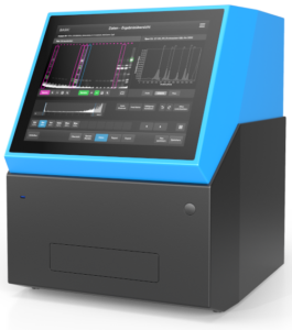
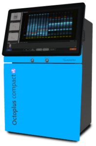
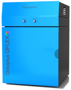
We cover a wide range of applications with our camera-based detection systems. You can choose between three different models:


ORCA Analyzer

  • • developed for routine diagnostics (1D gels)
  • • highly sensitive fluorescence detection (RGB)
  • • samples detectable via barcode reader
  • • automated lane and band detection incl. analysis
  • • automated exports and reports directly into the desired directory


Learn more...


Octoplus Compact SPL

  • • for research and routine use (for 1D gels and blots)
  • • modular system with modular filter configuration for fluorescence

(RGB + IR)

  • • highly sensitive ECL detection
  • • optional with UV (DNA/RNA) and white light transilluminator (Vis)
  • • manual focus control for maximum sharp bands
  • • with integrated SPL software for Western blot normalizations
  • • with integrated touch screen or separate PC


Learn more...


Octoplus QPLEX

  • • for research and routine use (for 1D/2D gels and blots)
  • • largest homogeneously illuminated imaging area on the market

(25 x 20 cm)

  • • modular system with flexible filter configuration for fluorescence

(RGB + IR)

  • • highly sensitive ECL detection
  • • optional with UV (DNA/RNA) and white light transilluminator (Vis)
  • • manual focus control for maximum sharp bands/spots
  • • in vivo imaging (plants/small animals) possible


Learn more...


Posted in Allgemein | Comments Off on Imaging Systems for Fluorescence, ECL, UV & Vis

Maßgeschneiderte Protein-Services für schnelle Ergebnisse

NHD Protein und Proteomic Service

Auf der einen Seite eine anspruchsvolle Fragestellung über bekannte oder noch unbekannte Proteine. Auf der anderen Seite der Wunsch nach schnellen Ergebnissen.

Seit über 15 Jahren bietet die NH DyeAGNOSTICS GmbH (NHD) maßgeschneiderte Protein und Proteomic Services an. In einem breiten Leistungsspektrum spielt die Identifikation von differentiell regulierten und/ oder modifizierten Proteinen aus komplexen Proben eine wichtige Rolle.

Neben der Komplexizität ( z.T. über 1.500 verschiedenen Proteine) kann auch die Zusammensetzung oder Beschaffenheit einer Probe grundsätzlich eine große Herausforderung darstellen.

Hier unterstützt NHD mit seiner umfangreichen Geräteausstattung und der großen Erfahrung in der Protein-Analytik. Der Nachweis von (unbekannten) Proteinen bei Streß-toleranten Muscheln, rauchenden Mäusen, trainierten Muskeln oder schwitzenden Menschen, es sind immer maßgeschneiderte Protein Services, mit denen wir schnell zum Ziel kommen.

.

Etablierte Top-down-Proteinanalytik

Insbesondere bei  komplexen Proben sowie bei (posttranslational) modifizierten Proteinen und Isoformen ist die Top-Down Proteomik der ideale Ansatz. Dabei werden zunächst Proteine getrennt und charakterisiert. Erst dann erfolgt deren Identifikation und ggf. weitere Charakterisierung auf Peptidebene.

Um schnelle und valide Ergebnisse produzieren zu können, setzen wir in der Regel auf die leistungsfähigste Methode in der Trennung von intakten Proteinen, der 2D-Gel Analyse. Diese Methodik erfordert vor allem viel hands-on Erfahrung, aber die konnte NHD über die Jahre immer weiter ausbauen.

Erst nach dieser Trennung und der Auswahl von spannenden Protein-Kandidaten geht es auf Peptidebene weiter. Je nach Fragestellung nutzt NHD dazu verschiedene massenspektrometrische Methoden.

.

Publikationsreife Daten in kurzer Zeit

Wir identifizieren die spannenden Proteine für unsere Kunden aus ihren Proben. Und die können dann die wirklich interessante Forschung daran fortführen.

"Wir bearbeiten kleine Aufträge genauso wie große, umfangreiche Studien. Wenn das Probenmaterial nicht der limitierende Faktor ist, dann sind erste, publikationsreife Daten in Wochen bis wenigen Monaten die Regel." sagt Dr. Jana Heise, Leiterin der Protein Services der NH DyeAGNOSTICS GmbH in Halle (Saale).

.

.

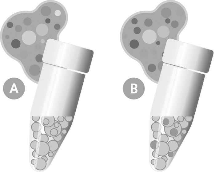
Bild zweier Proteinproben für eine vergleichende Proteom-Analytik

.

.

.

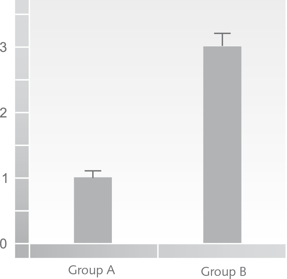
Bild eines Histogramms einer differentiellen Proteom-Studie

.

.

.

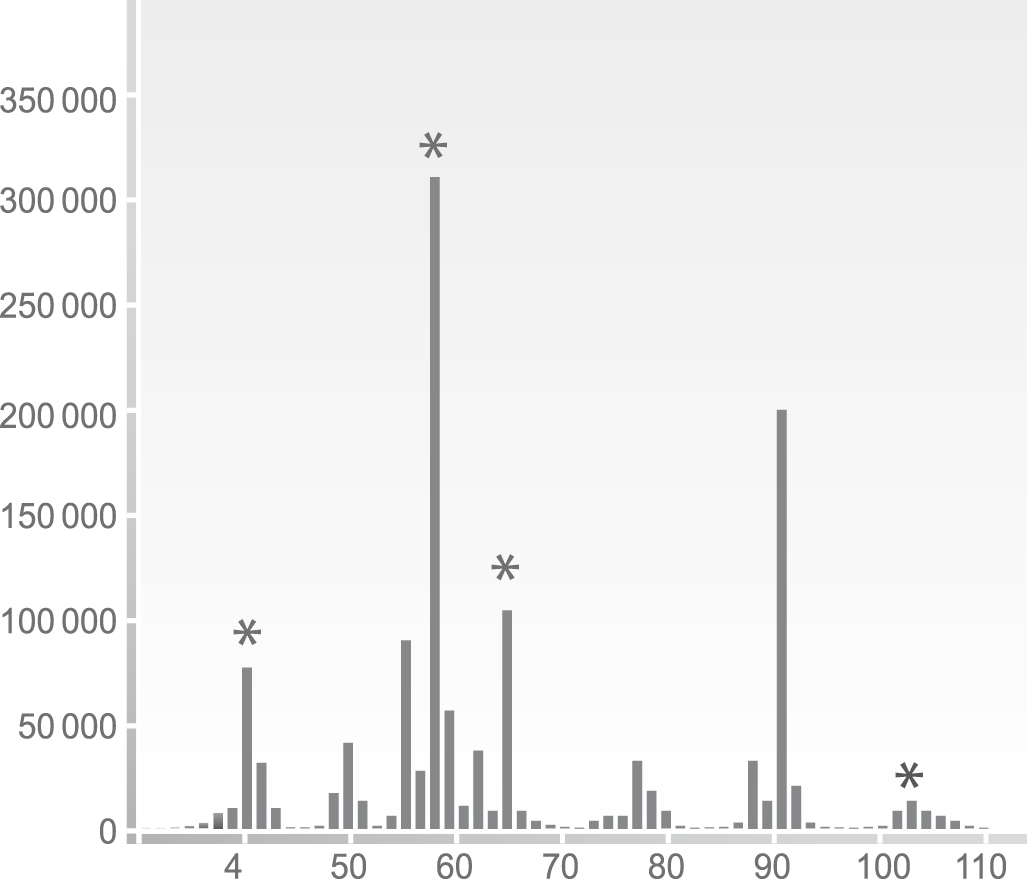
Bild eines massenspektrometrischen Fragmentierungsmuster eines Peptides

Posted in Allgemein | Tagged , , , , , , , , | Leave a comment

New VELUM Gels: Replacement for EXCEL gels

More reproducibility within your gel-based protein analytics?

VELUM Precast gels for horizontal gel electrophoresis are ready-to-use, so you do not need to prepare buffers. They are characterized by their easy handling (positioning in electrophoresis chamber / sample loading) and are therefore a perfect replacement for EXCEL gels.

Our portfolio includes gels for both 1D and 2D protein analytics (linear or gradient). In addition to SDS gels, we also offer IEF gels with a wide range of ph gradients. Further advantages of VELUM Precast gels are:

  • • optimized background reduction for fluorescence applications
  • • suitable for all protein staining applications (e.g. Coomassie, silver)
  • • individual gel composition possible upon request
  • • choice between different pocket sizes
  • • more reproducible running behavior
  • • even better selectivity
  • • new: ready-to-use
Posted in Allgemein | Comments Off on New VELUM Gels: Replacement for EXCEL gels

Western Blots: quantitative , valid results

The difficulties in obtaining Western But data

Western Blot analysis is based on a stringent work flow. Starting with the protein extraction, separation, transfer, antibody washing & incubation steps it finally come to the immunochemical detection of a specific protein.

In order to obtain valid, quantitative data, Western Blot experiments requires critical validation steps (Pillai-Kastoori L. et al., 2020).

.

Close monitoring of the sample

The patented SPL technology is based on the fluorescent labeling of the total protein after sample extraction. This speedy step enables the rapid but sensitive monitoring of the total protein within all further steps like separation (e.g. SDS-PAGE), transfer and target detection.

.

What do I need for using SPL?

Detection of SPL-labeled protein requires an adequate fluorescent imaging device. For Western Blots it allows for the co-detection of target and corresponding total protein of the sample.

Posted in Allgemein | Leave a comment