Western Blotting
 |
Octoplus QPLEXWestern Blot and Gel Doc ImagerIf you are looking for a robust device for reliable, high sensitivity Western Blot detection, have a closer look on the Octoplus compact SPL. |
||
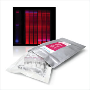 |
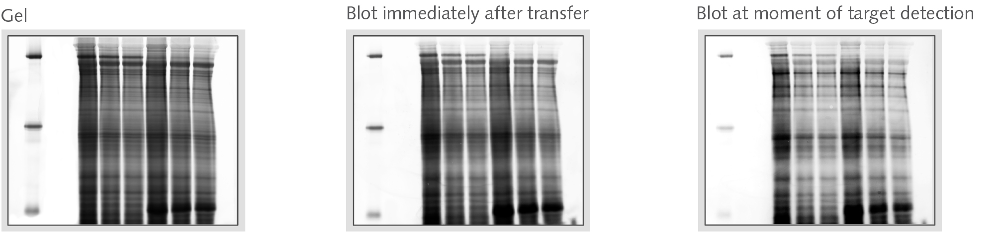
Smart Protein LayersKit for standardized total protein detectionSmart Protein Layers (SPL) is a patented standard-based technology for stain-free, quantitative analysis of total protein in protein gels and on Western blots at the point of time when the target is detected.SPL sets a novel standard for fast, reliable and standardized protein expression in gels and on blots. |
||
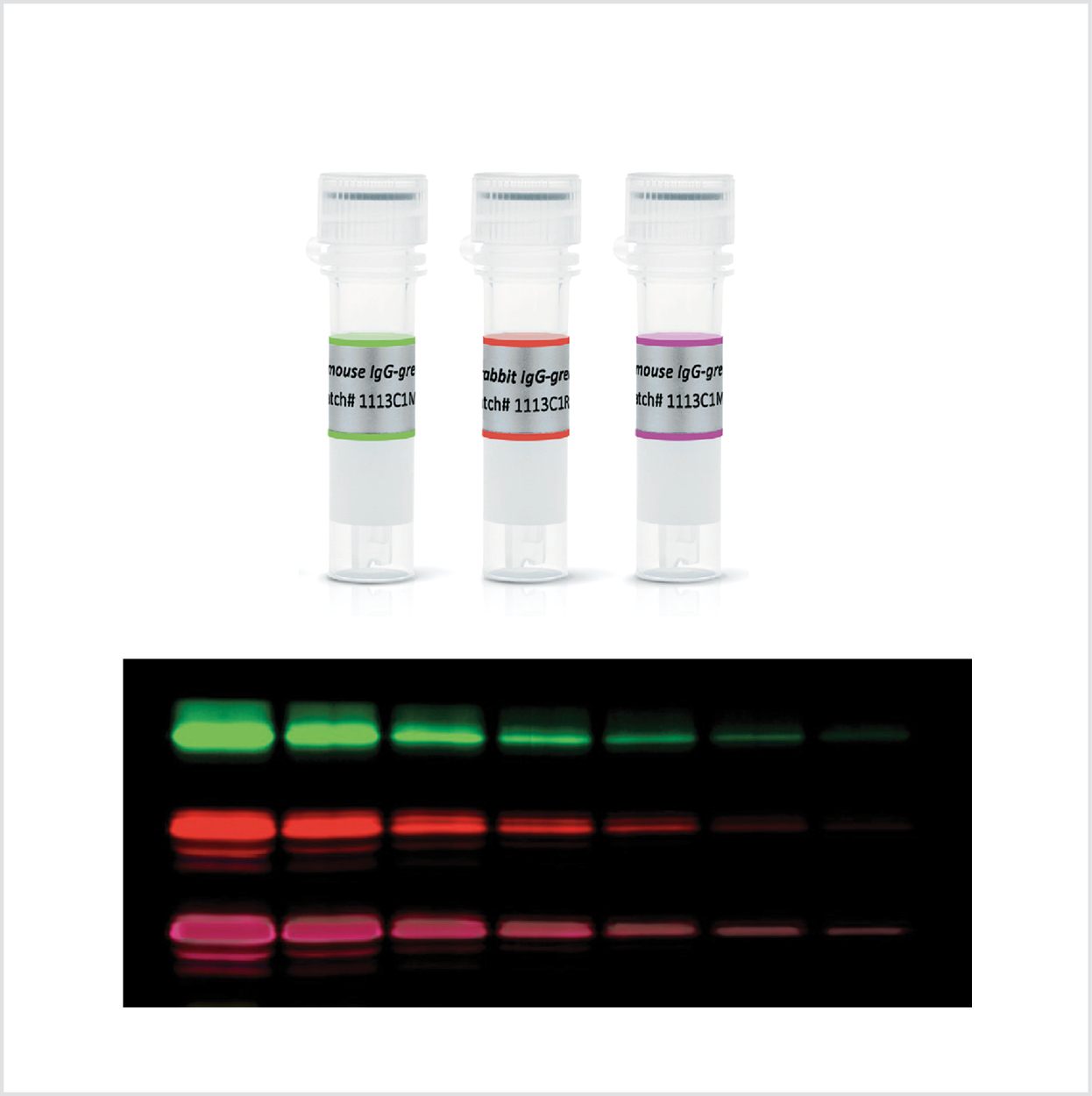 |
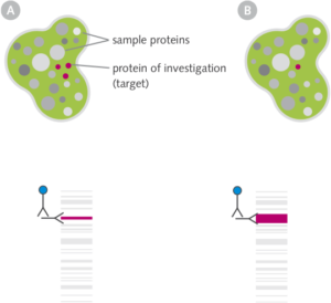
Fluorescent SecondariesDetection of target proteinsGenerally, fluorescent conjugated secondaries are the first choice for target protein detection on Western blots (if the target is not slightly above detection limit, than one must go for ECL).• Very easy handling • Signal stability of several months • Sensitivity in the higher pg range |
||
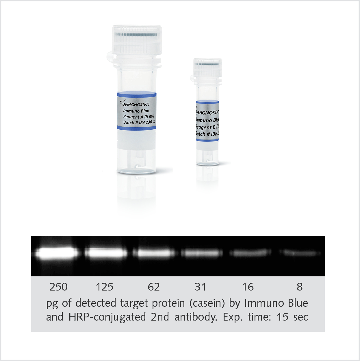 |
Immuno Blue HRP substrateDetection of the targetThe Immuno Blue HRP Fluorescence Substrate combines the high sensitivity of chemiluminescence with the signal stability and short detection time of fluorescent antibodies. Here, HRP-conjugated antibodies convert the Immuno Blue Substrate into a fluorescent precipitate that is stable over months. |
||
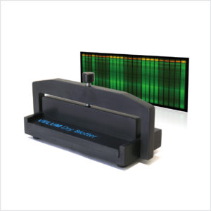 |
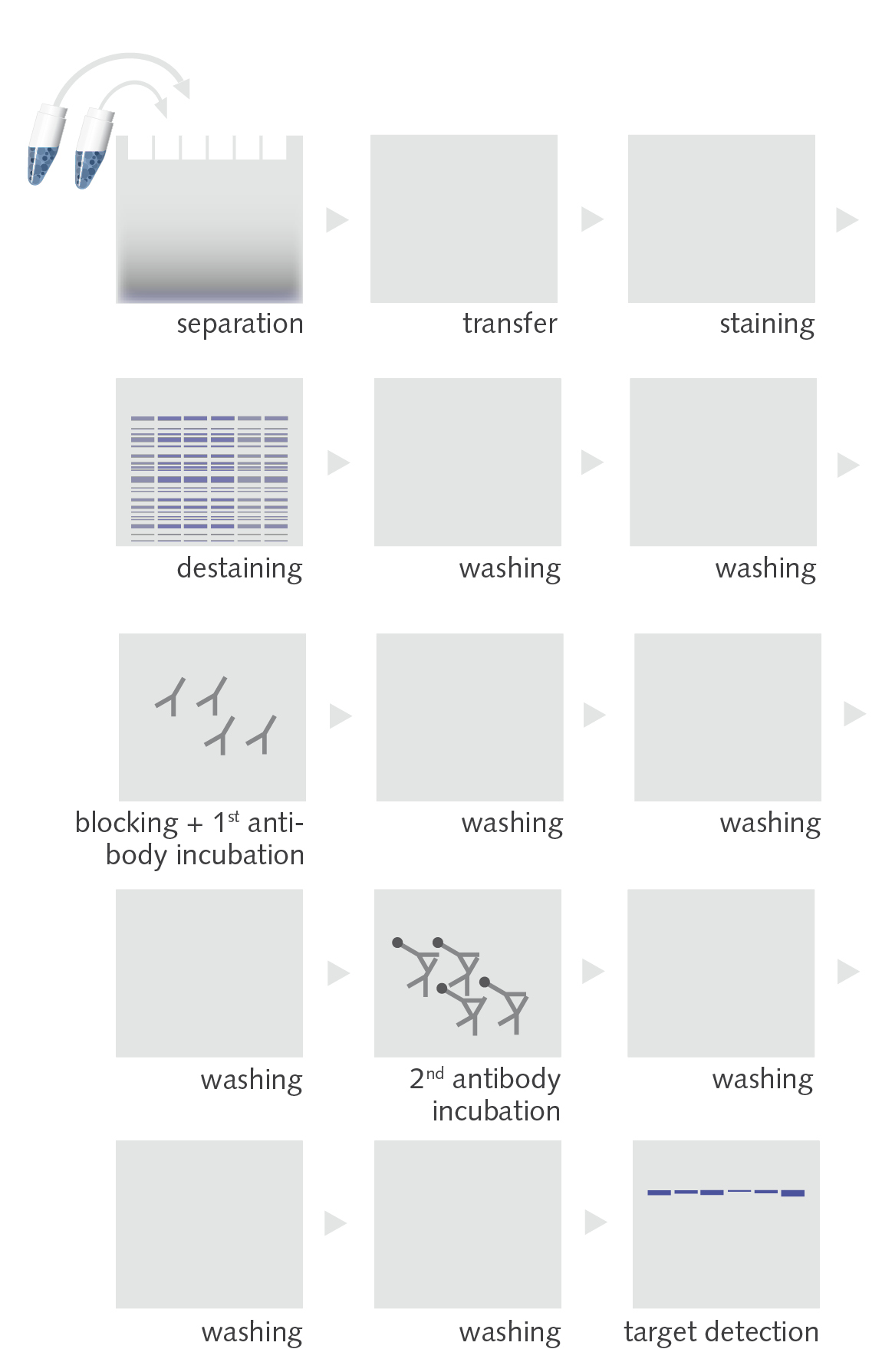
BlotterProtein transferThe BEO und VELUM Dry Blotter were originally designed for the transfer of proteins from plastic-backing supported precast gels like EXCEL or VELUM gels. However, their easy handling, no need of buffers and the high quality of the blots (e.g. no partial poor transfers results due to air bubbles) made them interesting for every user of blotting devices.Product page |
||
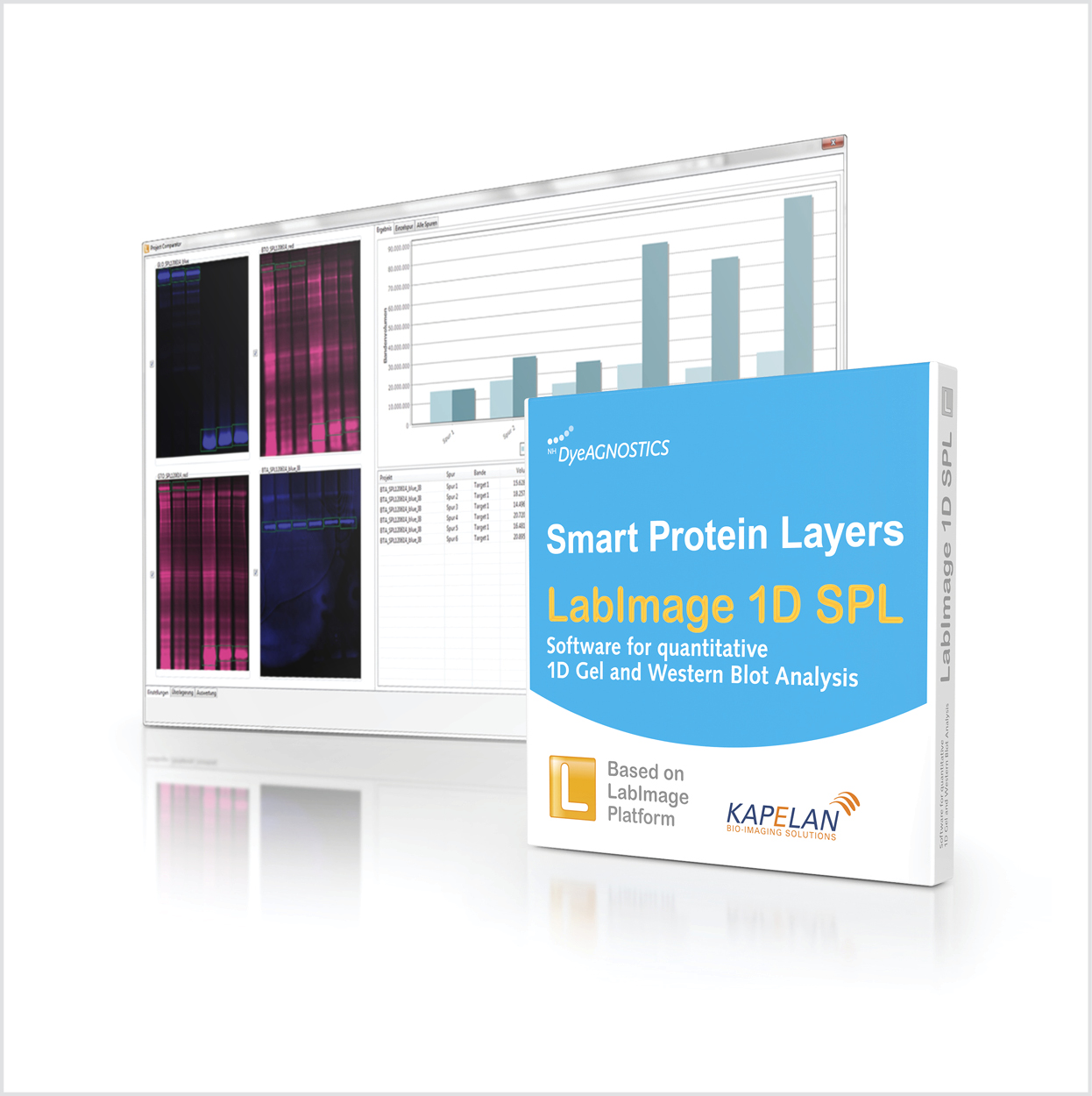 |
SPL LabImage 1D softwareFast and precise analysis of quantitative Western Blots using SPLThe SPL software allows for rapid analysis of quantitative Western Blots and 1D gels. The total protein and sample standards of the different sample are rapidly detected. Differences in protein content or amount of protein sample applied to the gel are monitored, calibrated and normalized. Target protein expression is automatically normalized to its corresponding sample total protein and sample standard.Product page |
||
.