
2D Protein Labeling Kits
Two-dimensional gel electrophoresis (2DE) is ideal for the analysis of intact proteins.In contrast to (peptide based)
LC-MS analysis, our high performing 2DE analyzes intact proteins which may include all information about:
- • post translational modifications such as phosphorylation, glycosylation or oxidation
- • splicing variants
- • protein isoforms
- • molecular weight
- • pI
We use our own fluorescence labeling technologies, so that we can offer you a wide portfolio of different 2D labeling kits for different purposes.


2DE Applications (selection)
- • Characterisation of a single proteome (protein pattern, PTM and isoform distribution): T-Rex 2D
- • Proteome profiling (differentiall analysis of two or more proteomes): (Refraction-2D, Saturn-2D)
- • Redox state analysis: Saturn-2D REDOX
- • Analysis of post-translational modifications (e.g. Phosphoryltion and Glycosylation): T-Rex 2D-PHOS and T-Rex 2D-GLYCO
- • 2D Western Blot Analysis: T-Rex 2D WB
- • Host Cell Protein Analysis: T-Rex 2D HCP
2D Precast Gels, 2D Electrophoresis Units & 2D Blotter
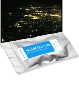

Velum Gold 2D Precast Gels
for high resolution 2D separation
.
2D Analysis Software and 2D Image Acquisition
.

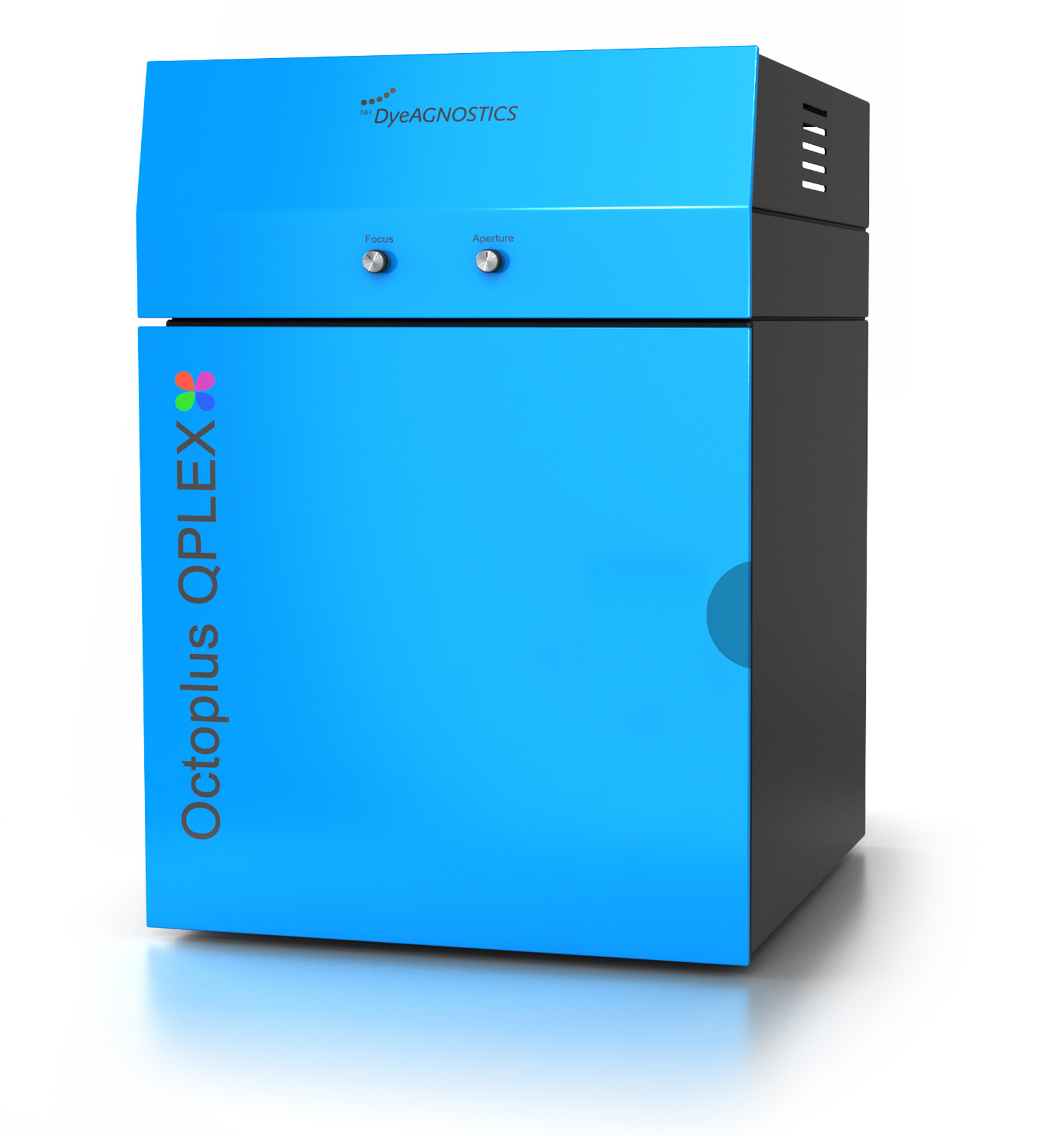
.
Two-dimensional gel electrophoresis
The principle of 2DE
The two-dimensional gel electrophoresis (2-D electrophoresis, 2-DE) is the most powerful method to separate complex protein mixtures.
It is based on the separation of intact proteins (not peptides) according to two molecular properties: the electric charge and the size of a protein.
The 2-D electrophoresis starts with the first-dimension separation - proteins are separated according to their electric charge, that is their isoelectric point. This step is called isoelectric focusing (IEF).
The IEF is (now a days) performed on a IEF strip, an acrylamide strip containing a pH gradient. When an electric potential is given to the IEF strip, the proteins will move along the strip to that point where their electric charge is 0 (neutral charge). After the IEF, the IEF strip now holds “a line of” the separated proteins according to their electrical charge.
After a step of equilibration the IEF strip is then applied to a SDS-PAGE gel in order to run the second dimension. This separation step now separates the now denatured proteins by their molecular weight.
.
2DE separation capacity
The protein separation capacity is given by
- a) the capacity of protein load of the IEF strip
- b) the separation area of the second dimension (the size of the SDS-PAGE gel).
For protein extracts from E. coli one should separate about 500 different proteins (e.g. detected by T-Rex fluorescent protein label) with 7 cm IEF strips and mini gel size SDS-PAGE (8 x 10 cm) and about 1200 (e.g. detected by T-Rex fluorescent protein label) proteins when using 24 cm strips and maxi size SDS-PAGE (27 x 22 cm).
.
Protein detection/ visualization
The number of proteins which can be detected is dependent on the method of protein visualization.
The general sensitivity limit is...
- • for Coomassie stains: a protein with an abundance of in the range of 100-10 ng can be detected.
- • for silver nitrate stains: a protein with an abundance of about 10-1 ng can be detected.
- • for fluorescence protein labels (minimal lysine labeling): proteins with an abundance of about 1 – 0.1 ng can be detected*.
- • for fluorescence protein labels (saturation cysteine labeling): proteins with an abundance of about 0.1 – 0.01 ng can be detected*.
* adequate fluorescence detection imaging device required
.
Differential Gel Electrophoresis/ Sample multiplexing
The usage of fluorescent protein labels is a strong benefit of 2D gel electrophoresis as the detection limit and dynamic range of the signal is much better and the analysis does not suffer from protein staining artifacts like background issues, difference in results due to room temperature, freshness of stains, etc… .
However, as proteins carrying a covalently bound fluorescence tag (label), they can be specifically detected. This allows to combine many samples to one 2DE analysis to allow for a precise quantification of protein expression and/or the accurate detection of post-translational protein modifications and to dramatically reduce the number of required 2D gels.