Western Blotting
 |
Octoplus compact SPLWestern Blot & Gel Dok ImagerWenn Sie ein robustes Gerät suchen, mit dem Sie in hoher Sensitivität und großer Zuverlässigkeit Western Blots durchführen können, das sollte dieses Gerät bei Ihnen in engster Auswahl stehen.Homogene und knackige Fluoreszenz von blau (ca. 500nm) bis Infrarot (ca. 770nm), sensitive Chemilumineszenz-Detektion, zahlreiche weitere Konfigurationsmöglichkeiten. Ausgelegt für tägliche Nutzung inkl. Praktikum. Empfohlen für die SPL-Technologie.Zur Produktseite |
||
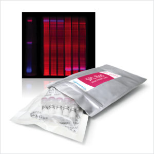 |
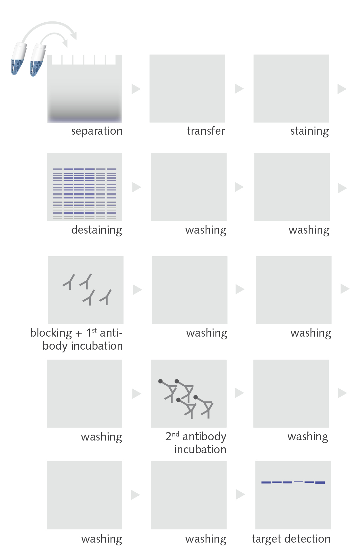
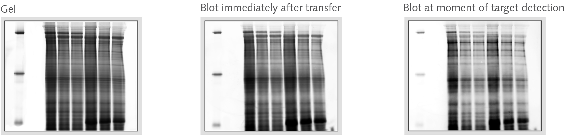
Smart Protein LayersKit zur Visualisierung des GesamtproteinsSmart Protein Layers (SPL) ist die patentierte, standard-basierte Technologie für die färbungsfreie, quantitative Analyse von Protein 1D-Gelen und Western Blots. SPL kombiniert dabei den schnellen und hochsensitiven Proteinnachweis mit einer neuen Qualität in punkto Normalisierung, Standardisierung und Quantifizierung.Zur Produktseite |
||
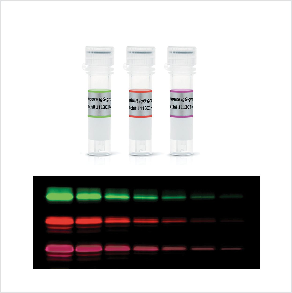 |
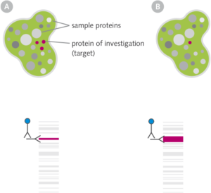
Fluoreszente Sekundär-AntikörperDetektion von TargetproteinenWenn Ihr im Blot nachzuweisendes Protein nicht knapp über dem Rauschen liegt (dann geht wirklich nur Chemilumineszenz), sollten fluoreszierende Antikörper die Nachweismethode der Wahl sein. Rote und infrarote Secondaries bieten grundsätzlich dabei die höchste Sensitivität (da das beste Signal-zu-Rausch-Verhalten). |
||
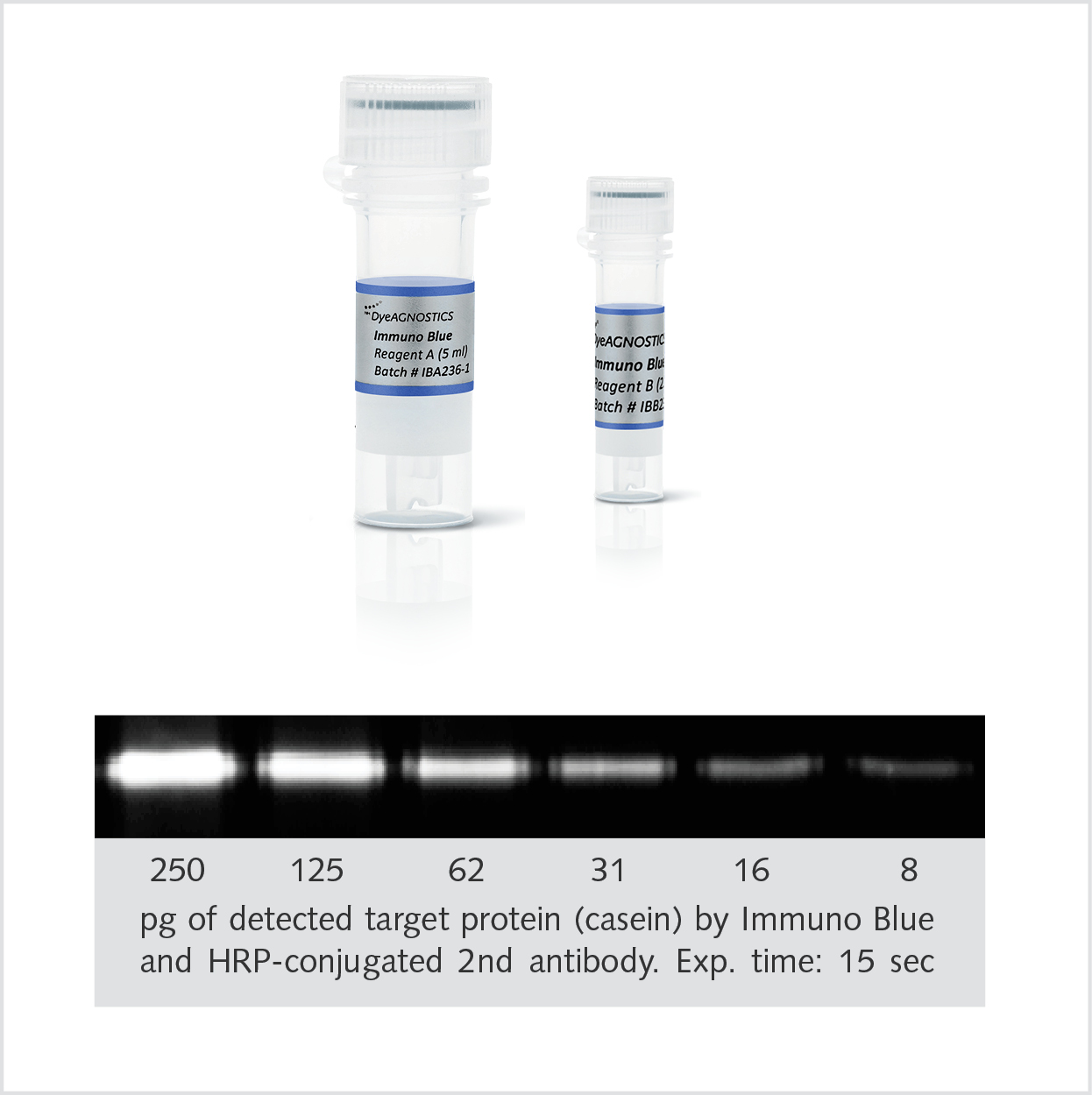 |
Immuno Blue HRP-SubstratDetektion des TargetsDas Immuno Blue HRP Fluorescence Substrate vereint die hohe Sensitivität der Chemilumineszenz mit der Signalstabilität und der kurzen Detektionszeit von fluoreszierenden Antikörpern. Hierbei wandeln HRP-konjugierte Antikörper das Immuno Blue Substrate in ein über Monate stabil fluoreszierendes Präzipitat um. |
||
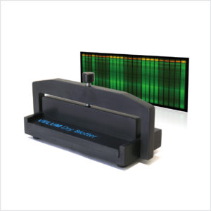 |
BlotterProtein TransferUnsere Blotterserie, der BEO Dry Blotter und der VELUM Dry Blotter, wurden für den Proteintransfer aus Foliengelen (z.B. EXCEL oder VELUM Gelen) entwickelt. Ihre einfache Bedienung ohne Puffer sowie die hohe Qualität der Blots (z.B. keine Luftblasen) machen diese einfachen Blotter aber auch zum beliebten Werkzeug für jede Art von Gel (Protein/ DNA / RNA).. Zur Produktseite |
||
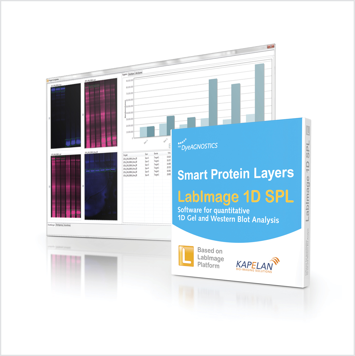 |
SPL LabImage 1D SoftwareSchnelle und präzise Auswertung von qWB AnalysenUm vertrauenswürdige quantitative Western Blots durchführen zu können, bedarf es der Normalisierung des detektierbaren Targets im Verhältnis zum Gesamtprotein auf dem Blot (natürlich zu diesem Zeitpunkt, da Proteine ja nicht kovalent an der Membran gebunden sind). Die SPL LabImage 1D Software berechnet mit einem Klick die Verhältnisse von Target und korrespondierendem Gesamtprotein pro Spur und über alle Spuren (Proben). |
||
.