
2D Protein Labeling Kits
Die zweidimensionale Gelelektrophorese (2DE) eignet sich ideal für die Analytik von intakten Proteinen. Die Proteine werden in zwei Dimensionen aufgetrennt, erstens nach dem isoelektrischen Punkt (pI) und zweitens nach ihrem Molekulargewicht. Dies resultiert in einer äußerst hohen Trennungskapazität von bis zu 2000 Proteinen in komplexen Proteomen. Im Gegensatz zu MS-basierten Analysen bleiben die Proteine während der 2DE-Analyse intakt und werden nicht zu Peptiden verdaut. Somit tragen die Proteine in der 2DE noch alle Informationen wie:
- • Post-translationale Modifikationen wie Phosphorylierungen, Glykosylierungen, Oxidationen
- • Protein Splicing-Varianten
- • Protein-Isoformen
- • Molekulargewicht
- • pI-Wert
Wir nutzen dabei unsere eigenen Fluoreszenz-Labeling-Technologien, sodass wir Ihnen ein breites Portfolio an unterschieldichen 2D Labeling Kits für verschiedene Fragestellungen anbieten können.


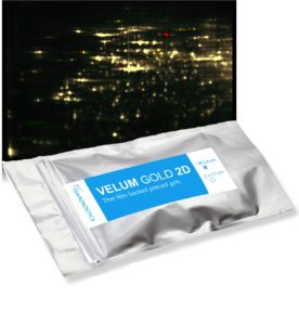
T-Rex
für hochsensitive, färbungsfreie Proteinvisualisierung
und 2D Western
2DE Anwendungen und die Analyse von intakten Proteinen (Auswahl)
- • Charakterisierung eines Proteoms (Protein-Muster, PTM- und Proteinisoform-Verteilung): T-Rex 2D
- • Proteom-Profiling (differentieller Vergleich mehrerer Proteome):
- • Redox-Status-Analysen: Saturn-2D REDOX
- • Untersuchungen von post-translationalen Modifikationen (Phosphorylierungen und Glykosylierungen):
- • 2D Western Blot Analysen: T-Rex 2D WB
- • Host cell protein analysis: T-Rex 2D HCP
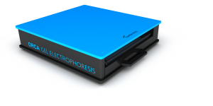
2D Fertiggele, 2D Elektrophorese-Systeme und 2D Blotter
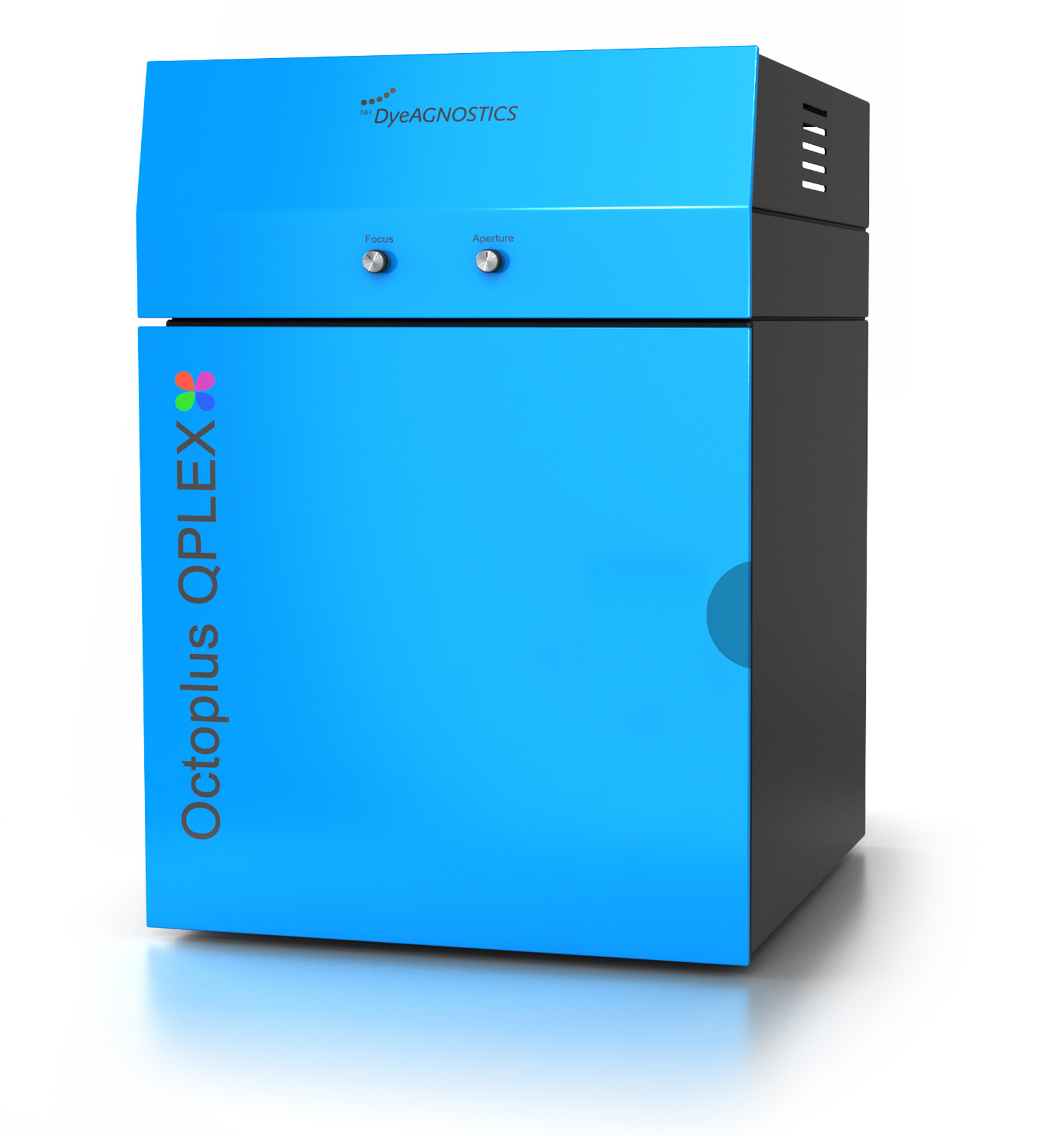
2D Auswertungssoftware und 2D Imaging
.

.
Two-dimensional gel electrophoresis
The principle of 2DE
The two-dimensional gel electrophoresis (2-D electrophoresis, 2-DE) is the most powerful method to separate complex protein mixtures.
It is based on the separation of intact proteins (not peptides) according to two molecular properties: the electric charge and the size of a protein.
The 2-D electrophoresis starts with the first-dimension separation - proteins are separated according to their electric charge, that is their isoelectric point. This step is called isoelectric focusing (IEF).
The IEF is (now a days) performed on a IEF strip, an acrylamide strip containing a pH gradient. When an electric potential is given to the IEF strip, the proteins will move along the strip to that point where their electric charge is 0 (neutral charge). After the IEF, the IEF strip now holds “a line of” the separated proteins according to their electrical charge.
After a step of equilibration the IEF strip is then applied to a SDS-PAGE gel in order to run the second dimension. This separation step now separates the now denatured proteins by their molecular weight.
.
2DE separation capacity
The protein separation capacity is given by
- a) the capacity of protein load of the IEF strip
- b) the separation area of the second dimension (the size of the SDS-PAGE gel).
For protein extracts from E. coli one should separate about 500 different proteins (e.g. detected by T-Rex fluorescent protein label) with 7 cm IEF strips and mini gel size SDS-PAGE (8 x 10 cm) and about 1200 (e.g. detected by T-Rex fluorescent protein label) proteins when using 24 cm strips and maxi size SDS-PAGE (27 x 22 cm).
.
Protein detection/ visualization
The number of proteins which can be detected is dependent on the method of protein visualization.
The general sensitivity limit is...
- • for Coomassie stains: a protein with an abundance of in the range of 100-10 ng can be detected.
- • for silver nitrate stains: a protein with an abundance of about 10-1 ng can be detected.
- • for fluorescence protein labels (minimal lysine labeling): proteins with an abundance of about 1 – 0.1 ng can be detected*.
- • for fluorescence protein labels (saturation cysteine labeling): proteins with an abundance of about 0.1 – 0.01 ng can be detected*.
* adequate fluorescence detection imaging device required
.
Differential Gel Electrophoresis/ Sample multiplexing
The usage of fluorescent protein labels is a strong benefit of 2D gel electrophoresis as the detection limit and dynamic range of the signal is much better and the analysis does not suffer from protein staining artifacts like background issues, difference in results due to room temperature, freshness of stains, etc… .
However, as proteins carrying a covalently bound fluorescence tag (label), they can be specifically detected. This allows to combine many samples to one 2DE analysis to allow for a precise quantification of protein expression and/or the accurate detection of post-translational protein modifications and to dramatically reduce the number of required 2D gels.