
.
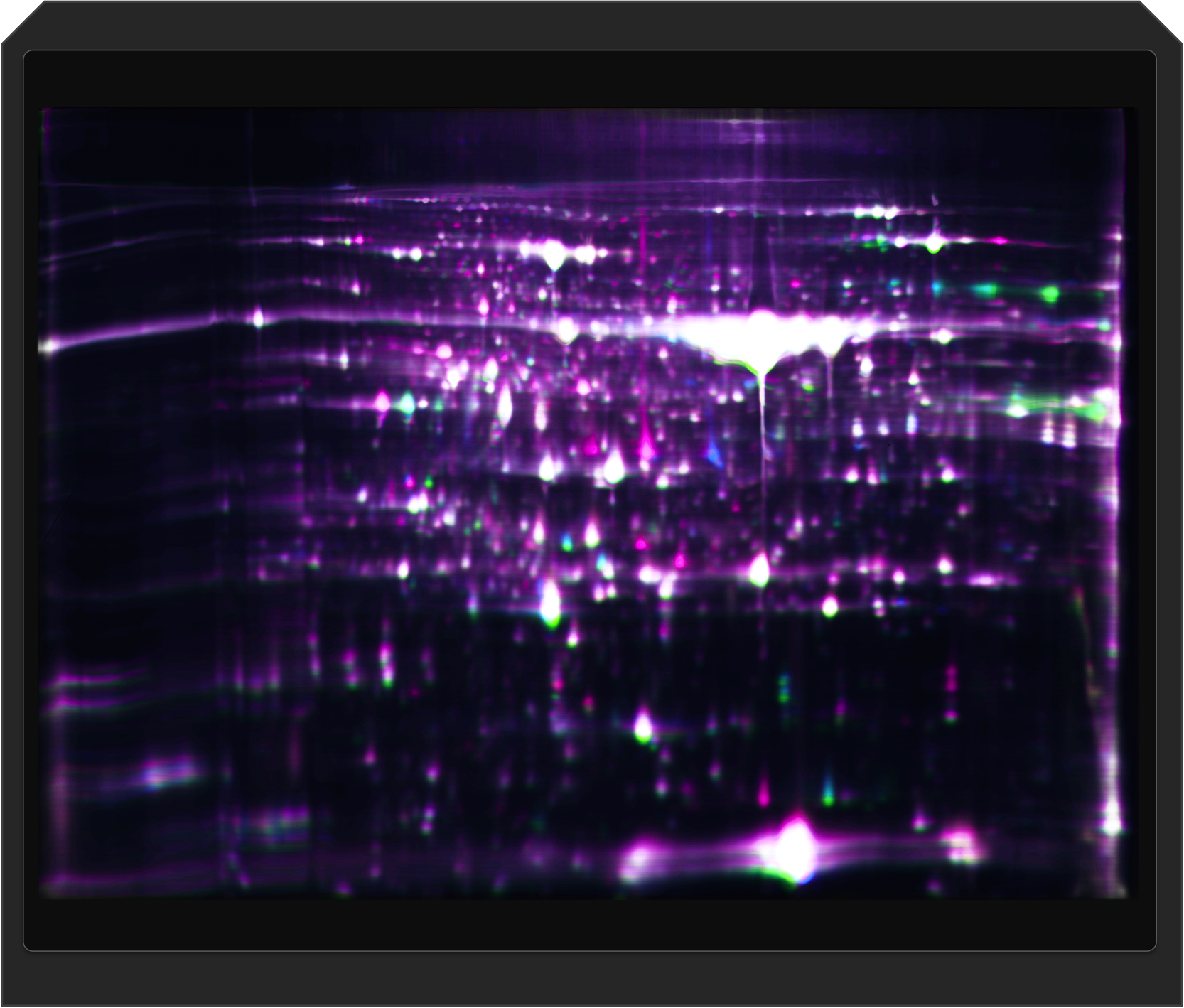
Refraction-2D/2D-DIGE/HCP
.
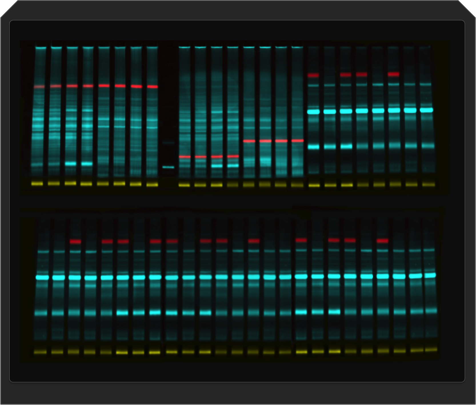
RGB+NIR Fluorescence/Chemiluminescence Western Blot
Spezifikationen
 |
homogene Multiplex-Fluoreszenz-Detektion ("publication level)" über eine Fläche von 20 x 25 cm |
 |
schnelle Fluoreszenz-Bildaufnahme
z.B. SPL Western Blots: 0.2 - 1.0 sec., VELUM Gold 1D Gel: ca. 1-2 sec. Refraction-2D pro Kanal: 10-30 sec. |
 |
leistungsstarke RGB- Fluoreszenz u.a. für
G-Dye100, G-Dye200, G-Dye300, Cy2, Cy3, Cy5, ... |
 |
leistungsstarke NIR- Fluoreszenz u.a. für
G-Dye400, LiCOR CW800, ... |
 |
leistungsstarke ECL Detektion |
 |
sensitive Coomassie-Detektion
(über rote Power-Fluoreszenz) |
 |
Remote- und Hands-on Unterstützung |
 |
geringster Wartungsaufwand |
 |
Entwickelt und hergestellt in Deutschland |
Übersicht Anwendungen (Beispiele)
| Multiplex Fluoreszenz | |
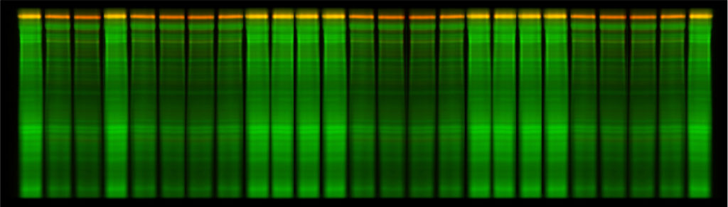
| quantitative/qualitative
1D Gele Größe bis zu 250 x 120 mm |
 |
| quantitative/qualitative
Western Blots Größe bis zu 250 x 120 mm |
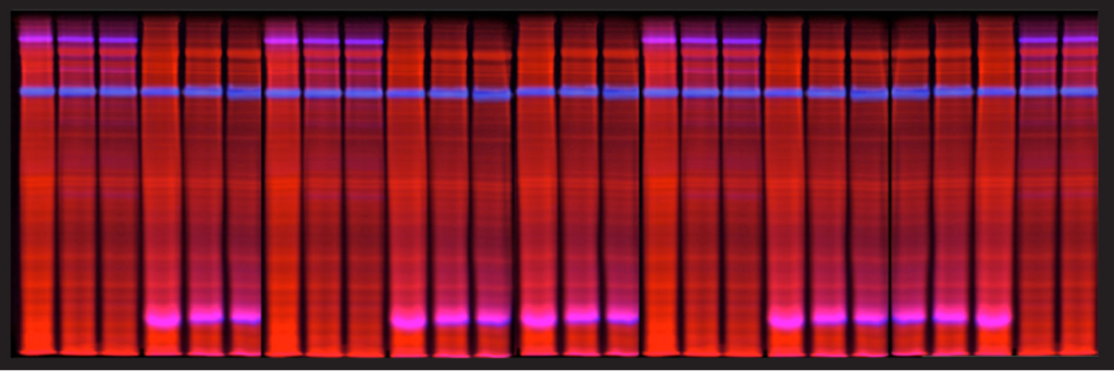 |
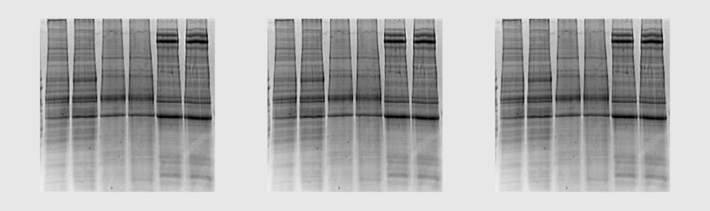
| quantitative/qualitative
Refraction-2D/2D-DIGE Gele und Western Blots z.B. für 24cm IEF-Streifen |
 |
| Chemilumineszenz | |
| ECL Detektion wichtig z.B. bei schwach-abundanten Targets | 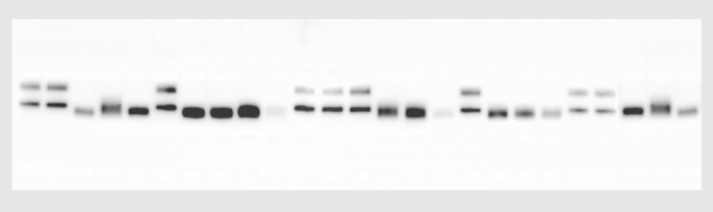 |
| Vis-Färbungen | |
| Coomassie-
Färbung (Detektion über rote Power-Fluoreszenz) |
 |

Support
Sehr gerne geben wir Ihnen weitere Auskünfte.
Rufen Sie uns doch an unter 0345 -2799 6413 (Mo - Fr 9.00- 17.00h)
oder schicken Sie uns eine kurze Email an info dyeagnostics.com.
.
